Medical imaging
At the Bergschenhoek location, radiographs can be taken, both 'normal' radiographs and dental radiographs (during dental treatment).
What are radiographs?
An X-ray uses X-rays to take a picture of the inside of the body. Certain tissues in the body block the X-rays, while others let them through easily. This makes it possible to see different structures in the body on an X-ray.
Why would your pet need X-rays?
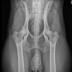
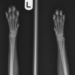
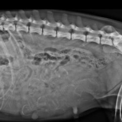
X-rays can be used to help diagnose various conditions and problems, ranging from bone fractures to problems with internal organs such as the lungs, liver and kidneys. X-rays are also used to produce official HD/ED radiographs to assess hip and elbow joints in certain breeds of dogs.

How does taking X-rays work?
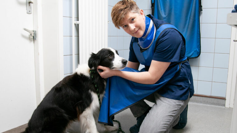
Taking radiographs is a non-invasive procedure in which your pet is placed on a table and possibly positioned in a certain 'posture'. Often this can be performed without anaesthesia, however in some cases a brief sedation may be required. The process is quick and comfortable for your pet, and our experienced veterinarians and para-veterinarians ensure that everything is done safely.
What happens after x-rays?
After the radiographs are taken, our vet will carefully assess the images (this is often done immediately) to make a diagnosis if necessary. If necessary, further investigations and/or treatments may be recommended based on the findings of the radiographs.